Dental X-Rays is also known as Dental Radiographs. It is a type of image of your teeth, with the help of that image, dentist evaluates and examine your oral health. it is an initial and essential part of dental treatment. Taking of X-Rays Help the dentist to find and Diagnosis problem at earlier stage before they become major problem

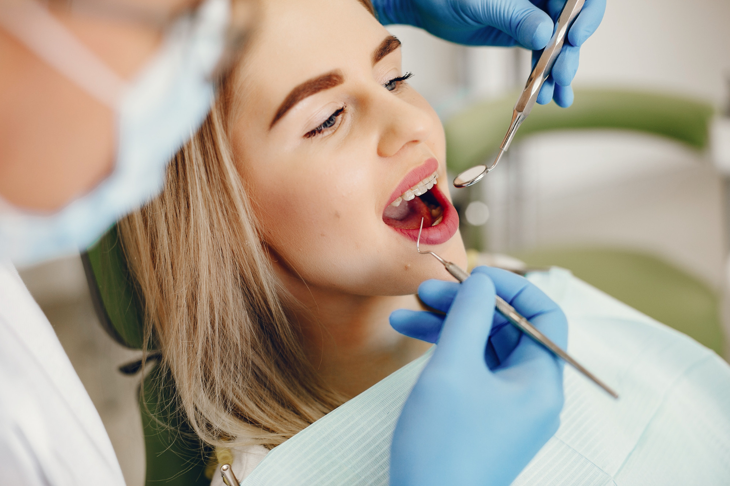
Dental X-rays is essential & useful tool for inspecting your oral health. Dentist can check your health by visual examination but there may be conditions that, some issues can only be detected with the help of X-rays Only.

Energy in the form of Rays is passed By X-ray Machine inside the mouth. Teeths, Bones & tissues of the mouth is very dense as compared to other organs, hence it is absorbed X-rays. infected part of the teeth or Bones or Gums or cheeks don’t absorb the X-rays, Hence it is look darker in the X-ray Film.

Dental X-Rays is useful to find abnormalities like:

• Cavities, Decay Between teeth and Fillings
• Periodontal Disease Impacted
• Missing and Cracked teeth
• Endodontic Problems
• Gum Disease, Different Types of Tumors

X-rays are mainly divided into two catagories

1.Intraoral X-rays
2.Extraoral X-Rays

1.Intraoral X-rays:

It is most commonly used type of X-rays. It gives the deep details about Cavities, Health of tooth root, and status of developing tooth.
Intraoral X-rays further Divided into 3 Parts

1.Bite-Wing X-rays:
This X-rays help dentist to find Decay between Back Teeth.

2.Periapical X-rays:This X-rays help dentist to find the entire lenth of each tooth from top to bottom,i.e from Crown to Root.

3.Occlusal X- rays:This X-rays Shows the arch of teeth of upper jaw or lower jaw. This X-ray help dentist to monitor the teeth development and placement.

1.Extraoral X-rays:

This X-ray gives proper details about teeth as well as Jaw and Skull. It gives less details than Intraoral X-ray. Hence they are usually not used.

Dental X-RAY OPG Advantages :

• Broad anatomic region is imaged.
• Low radiation dose.
• Convenient, easy and fast. The entire procedure takes 14 seconds exposure time.
• No overlapping of facial bones.

Indications :

• Evaluation of trauma, third molar, extensive pathology.
• Evaluation of tooth development especially mixed dentition analysis.
• Evaluation of development anomlies like cleft palate.
• Temporomandibular joint pathology, dislocation or fractures can be clearly visualized without overlap.
• Multiple dental/oral problems caused by tobacco/Pan masala chewing in elders & Chocolates in children are caught by a single X-ray.

Precuations While taking X-Ray-OPG :

You may be asked to remove jewellery, eyeglasses, and any metal objects that may obscure the images. You will be asked to stand with your face resting on a small shelf and to bite gently on a sterile mouth piece to steady your head. It is important to stay very still while the x-ray is taken.You will not feel any discomfort during the procedure.Pregenent women should Avoid X-ray

Lateral Cephalogram

A Lateral Cephalogram (or Lat Ceph) is an x-ray taken of the side of the face with very precise positioning, so that various measurements can be made to determine the current and future relationship of the top and bottom jaw (maxilla and mandible) and therefore assess the nature of a patient’s bite. This is particularly useful to plan any orthodontic treatment that may be necessary.

Advantages :

One of the biggest benefits of the lateral cephalometric x-ray is the advantage of “natural” head positioning. The lateral cephalometric x-ray uses a line known as the true vertical as an external reference point. Because the true vertical is generated by gravity and the head is in its normal positioning, a doctor or orthodontist can study the head as it would be positioned without any additional altering. This is why many refer to the lateral cephalometric x-ray as revealing the “natural” head positioning through the use of the true vertical.

Why take a lateral cephalometric x-ray?

Like other types of cephalometric x-rays, the lateral cephalometric x-ray is very helpful in dental and orthodontic treatment planning.

It can be used in many stages of treatment to determine what is the next best stage. However, the lateral cephalometric x-ray in particular is used by dentists and orthodontists because it can provide specific images of the mandible, or jaw, and its relationship to the maxilla, or cheek bone.

This can be useful in many different types of treatment plans, and it may be recommended as an analytical tool by your doctor. The difference between a lateral and horizontal cephalometric x-ray is that a lateral x-ray provide a photo of the side (lateral) view, while a horizontal x-ray provides a view of the top.

Procedure to take lateral cephalometric x-ray

The lateral cephalometric x-ray is a quick and painless procedure. It is taken in a Cephalostat, with the x-ray beam perpendicular to the patient’s sagittal plane. The beam most commonly enters on your right side, with the film cassette adjacent to your left side, so that your head is oriented to the right on the radiograph. While this is the most common type of lateral cephalometric x-ray, the reverse convention can also be used.

Uses of a lateral cephalometric x-ray

The lateral cephalometric x-ray will return a high-quality, 2D radiograph that the doctor can use to assess many different landmark points and planes of reference on your skull. The relationship of these to one another and to standard norms can then be quantified when determining appropriate steps in your treatment.

Open chat
1
Hello
Can we help you?